Chromosome number change
Aneuploidy
An increase or decrease in the number of an individual chromosome

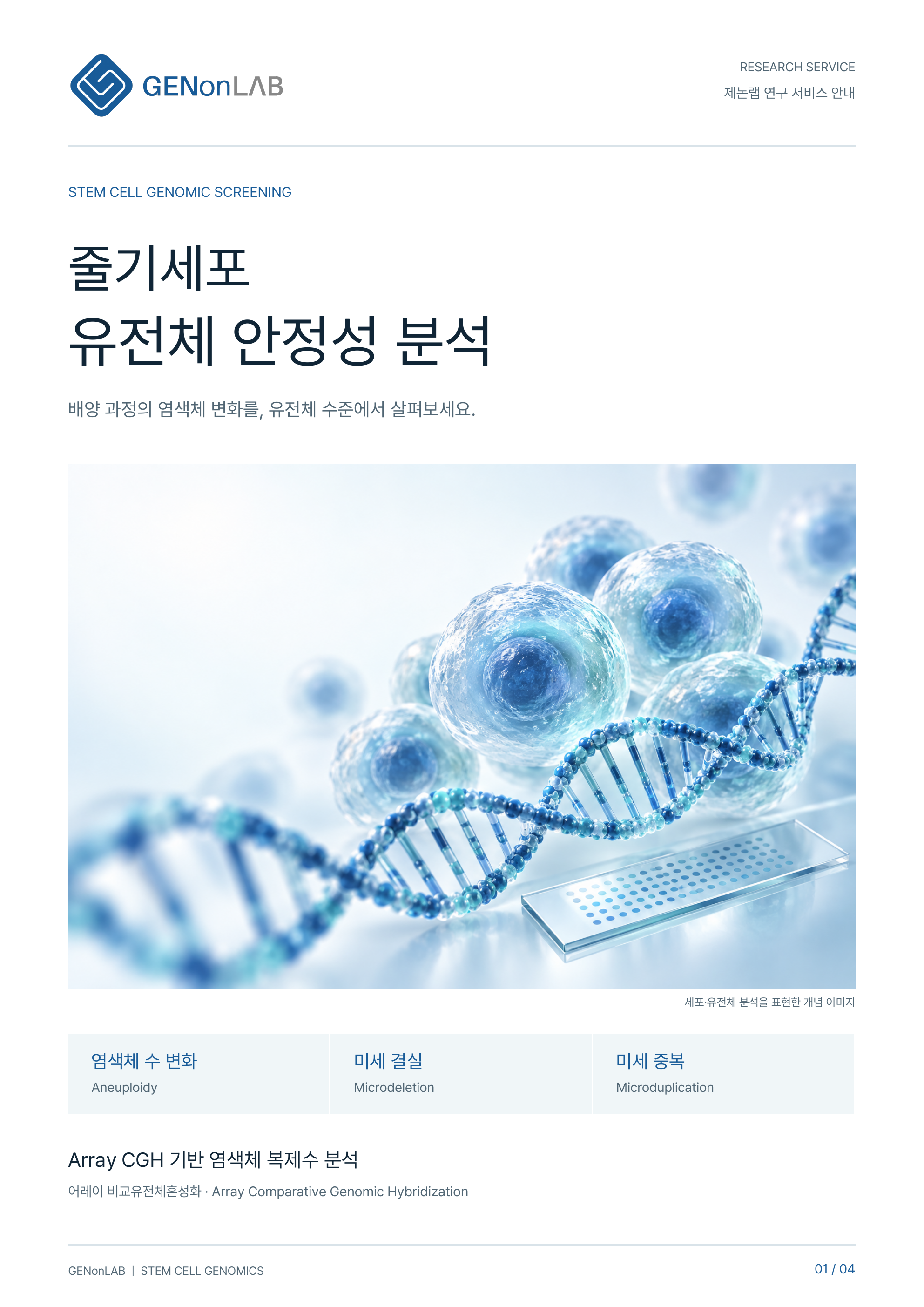
STEM CELL GENOMIC ANALYSIS
Your cells are growing well. Is their genome stable?
Chromosomal changes can arise during serial passaging. Assess research quality with genomic analysis suited to your purpose.
01 / WHY IT MATTERS
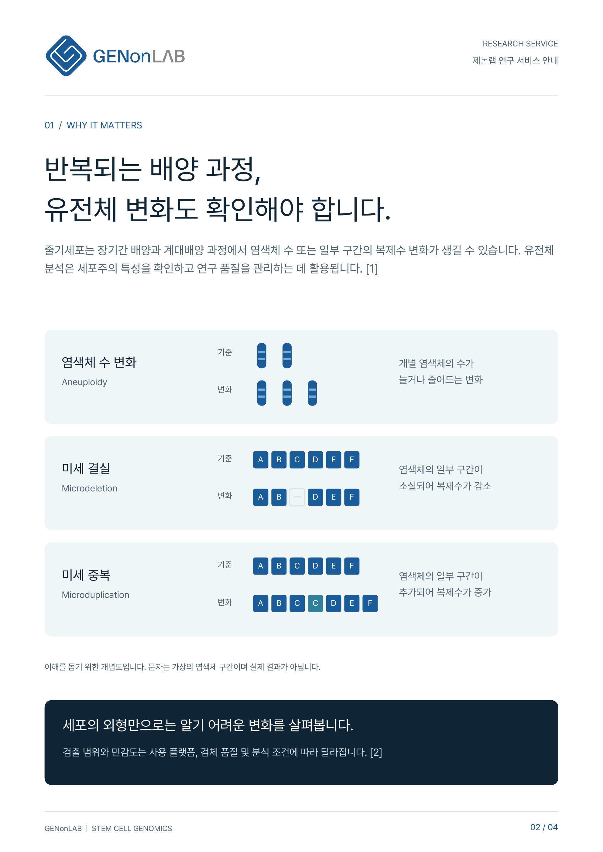
During long-term culture and passaging, stem cells may develop changes in chromosome number or segmental copy number. Genomic analysis helps characterize cell lines and manage research quality. [1]
Aneuploidy
An increase or decrease in the number of an individual chromosome
Microdeletion
Loss of a chromosome segment reduces its copy number
Microduplication
An extra chromosome segment increases its copy number
Conceptual diagrams for explanation only. Letters represent hypothetical chromosome segments, not actual results.
02 / ANALYSIS WORKFLOW
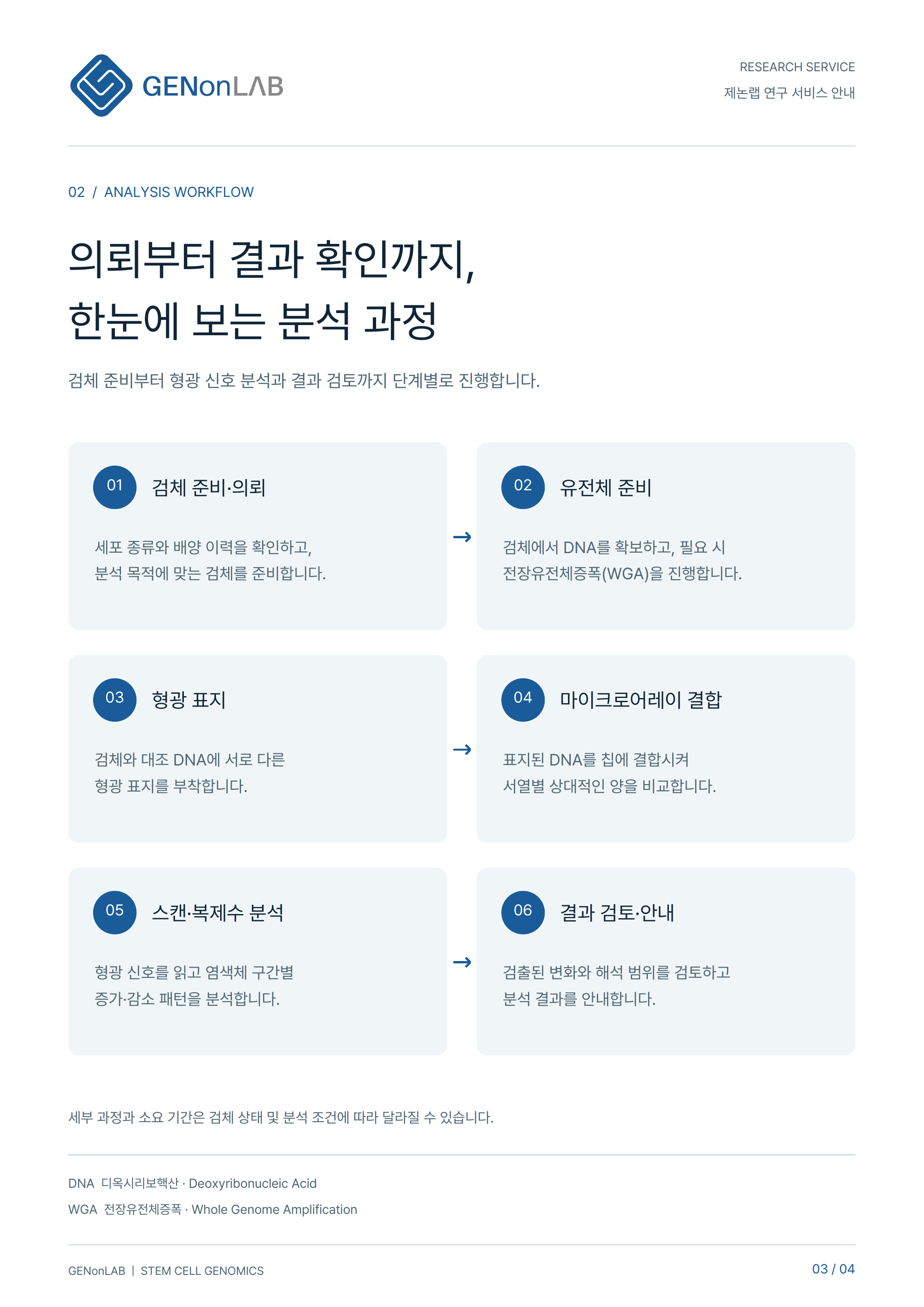
The workflow covers sample preparation, fluorescence signal analysis and result review.
Cell type and study purpose
Quantity, storage and shipment
Analysis after sample acceptance
Findings and interpretation scope
Confirm the cell type and culture history, and prepare a sample suited to the analysis objective.
Obtain DNA from the sample and perform whole genome amplification (WGA) if required.
Label the sample and reference DNA with different fluorophores.
Hybridize the labeled DNA to the array to compare relative amounts at each sequence region.
Read fluorescence signals and analyze gain and loss patterns across chromosome regions.
Review detected changes and interpretation limits, then report the results.
The detailed workflow and turnaround time may vary with sample condition and analysis requirements.
03 / INTERPRETATION
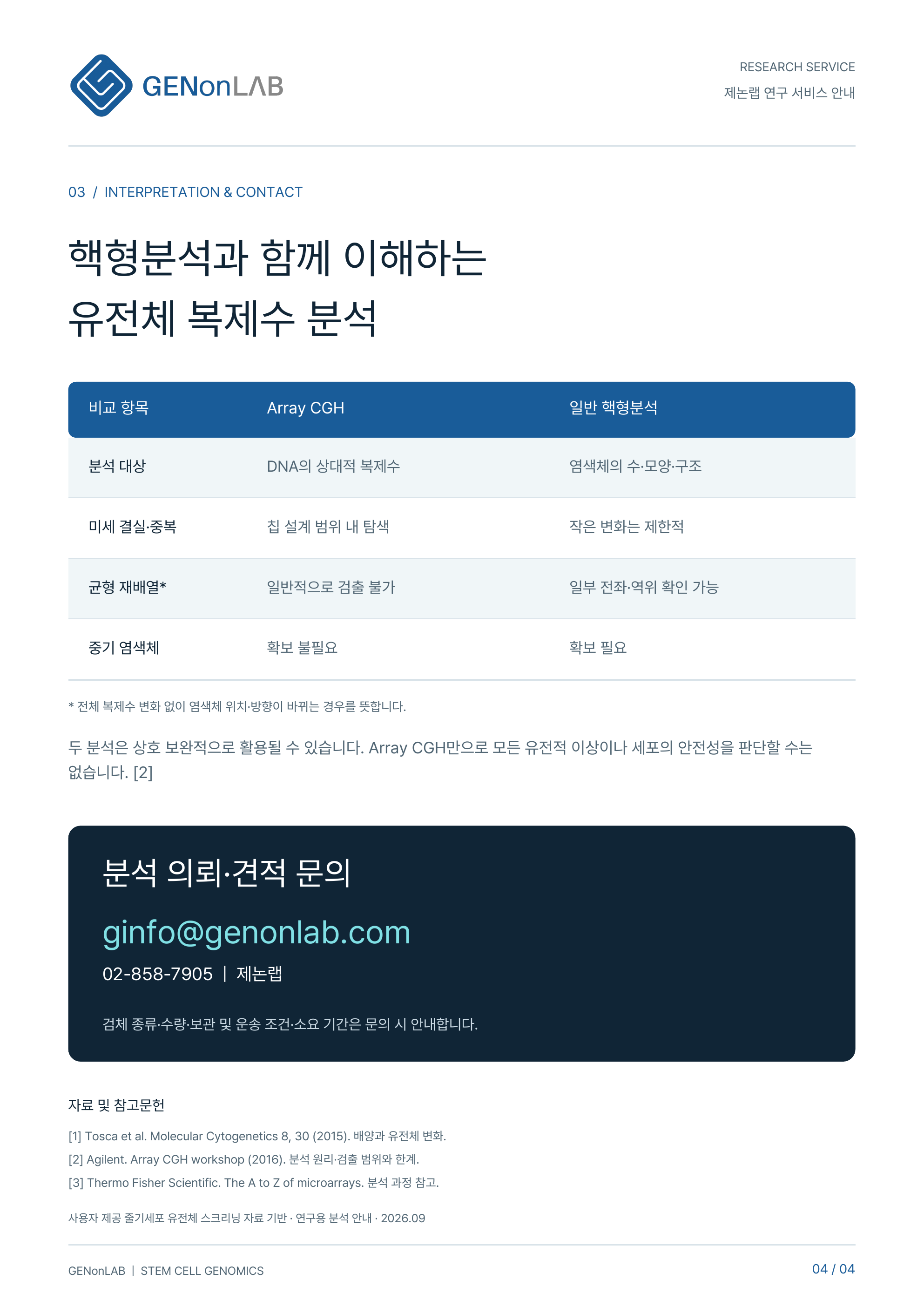
KARYOTYPE ANALYSIS
Examines chromosome number, morphology and structure, including translocations and inversions within the method’s detection range.
ARRAY CGH
Explores chromosome copy-number changes, including small deletions and duplications that may be difficult to identify with conventional karyotyping.
Array CGH generally does not detect balanced rearrangements without copy-number change. Discuss which methods and detection scopes suit your study.
Array CGH · Array Comparative Genomic Hybridization| Comparison | Array CGH | Conventional karyotyping |
|---|---|---|
| Analysis target | Relative DNA copy number | Chromosome number, shape and structure |
| Microdeletions & microduplications | Within the array design coverage | Limited for small changes |
| Balanced rearrangements* | Generally not detected | Some translocations and inversions can be detected |
| Metaphase chromosomes | Not required | Required |
* Changes in chromosome position or orientation without an overall change in copy number.
The two methods can be complementary. Array CGH alone cannot identify all genetic abnormalities or establish cell safety. [2]
BROCHURE
Two new promotional sheets · Open either image at full size. Artwork is in Korean; its key information is also available in the page text.
Research quality and three areas of focus
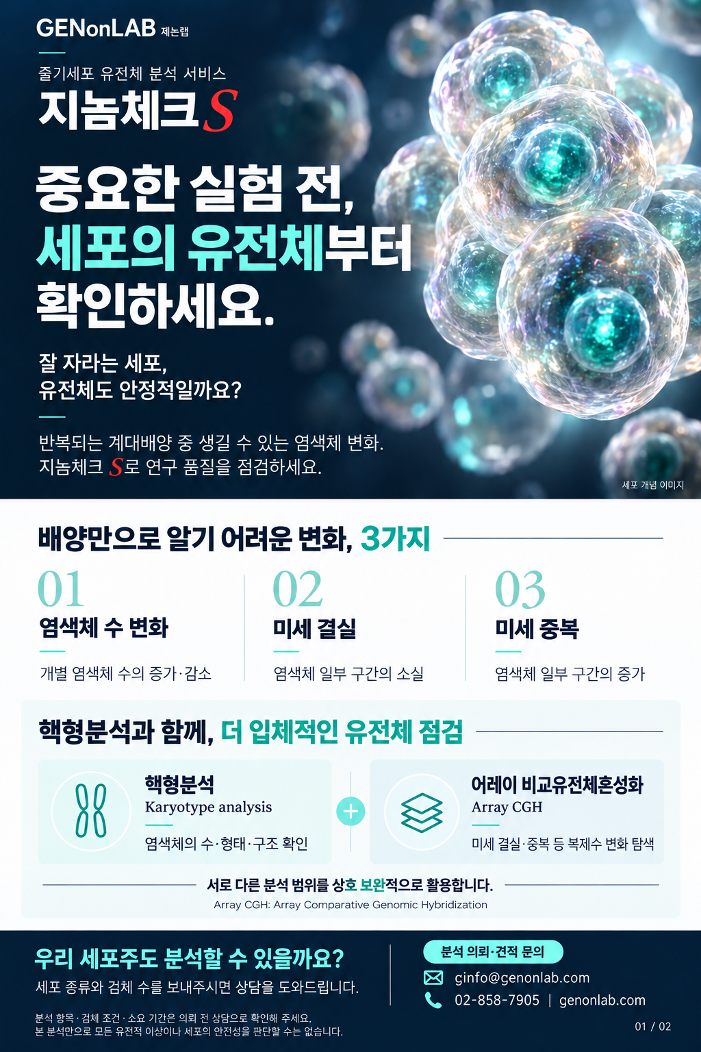
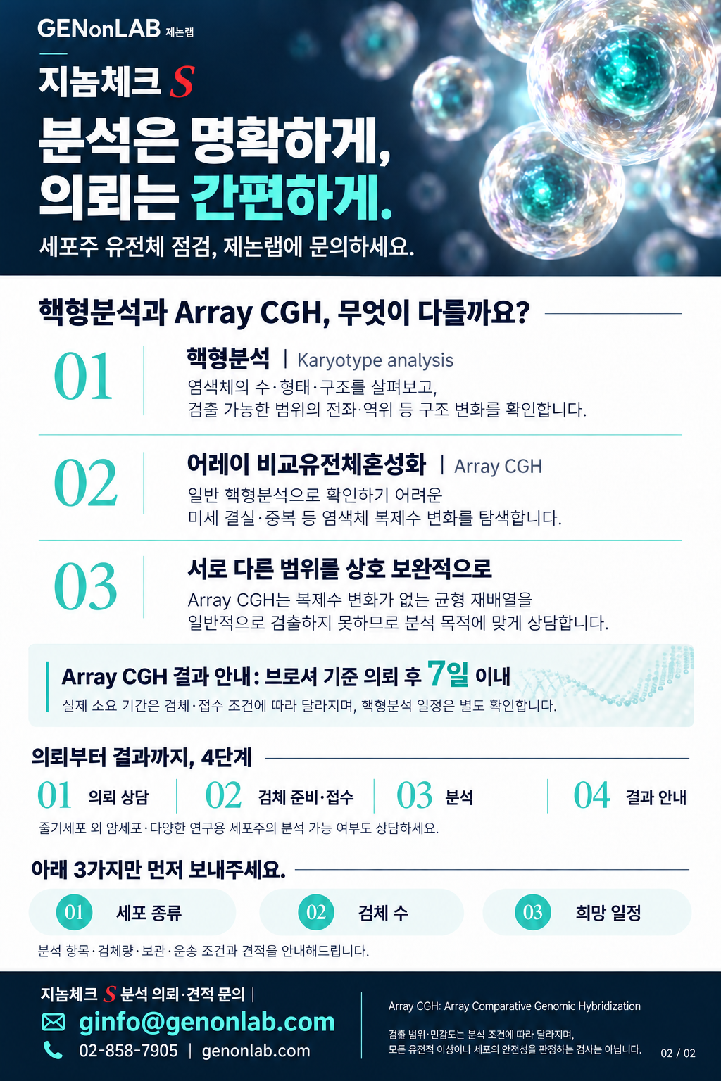
Karyotyping, Array CGH and submission guidance
REQUEST DOCUMENTS
Download and complete both forms below when requesting analysis.
Enter the information required for your test request.
Read the consent form and complete the required fields.
Click “Prepare analysis inquiry email” below, then manually attach your files to the email.
Email & contact detailsCONTACT GENonLAB
Start with three details.
Ask about feasibility for cancer cells and other research cell lines as well as stem cells.
Contact us to confirm sample type, quantity, storage, shipping conditions and turnaround time.
Complete the test request and genetic testing consent forms, then attach photographs or scans to your request email.
Include the cell type, analysis objective and requested schedule to help us review your request.
The inquiry button opens your email app. If it does not open, copy the address above and send your inquiry using your preferred email service.
Genomic analysis for stem cell research · September 2026